Anterior cruciate ligament and injuries (part-1)
Anterior cruciate ligament and injuries
The following article provides in-depth information about anterior cruciate ligament anatomy, injuries and its physiotherapy management.
One of the most common knee injuries is an anterior cruciate ligament (ACL) sprain, or tear. Athletes who participate in high demand sports like soccer, football, and basketball are more likely to injure their ACL.
Anatomy
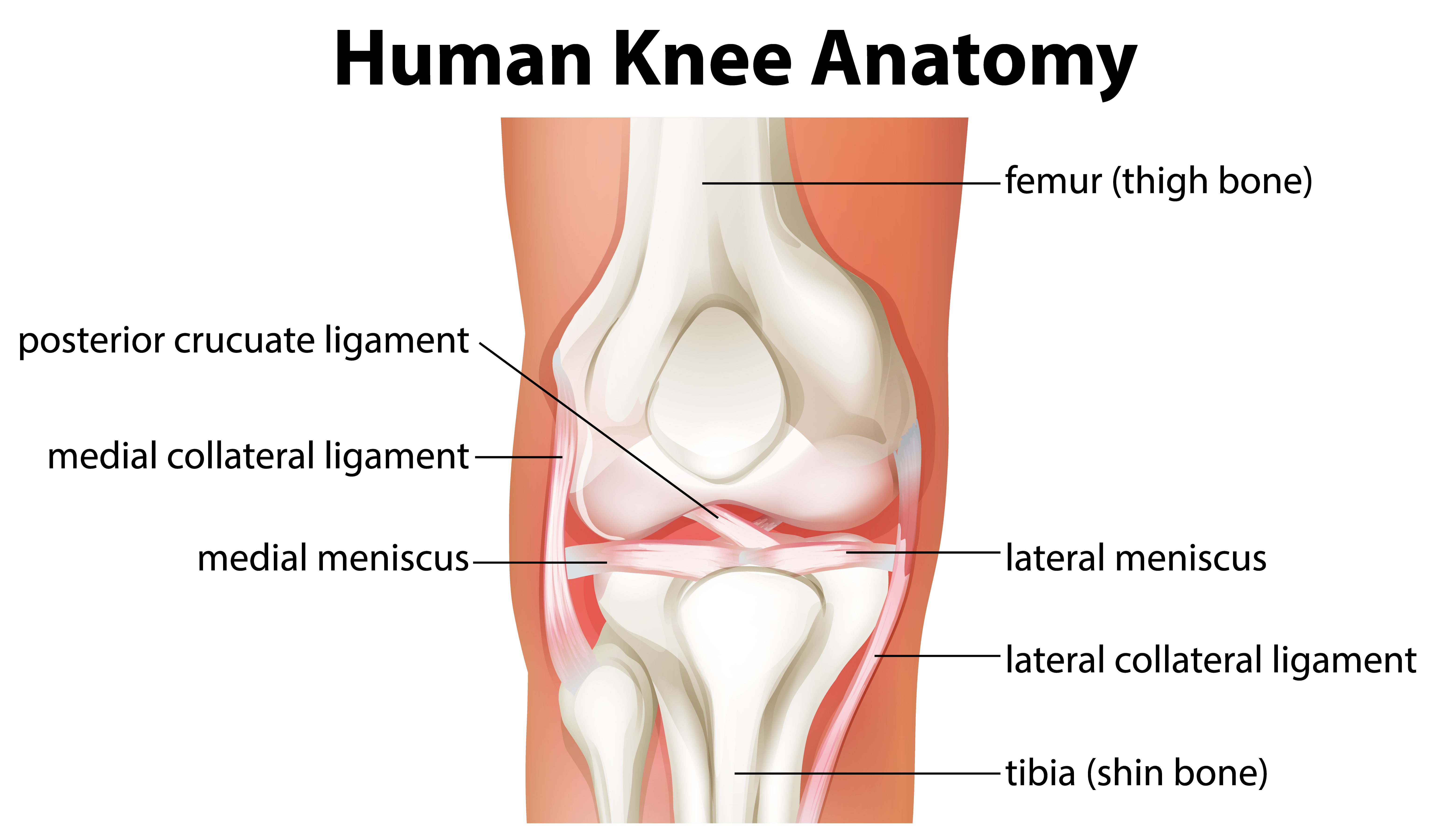
knee joint if form by union of three bones: femur (thigh), tibia(shin), and patella (kneecap). Patella is situated in front of the leg to provide protection. Generally, bones are connected to other bodes by the help of ligaments. There are 4 primary ligaments working as a strong rope to hold knee joint together and provide stability while its moving.
Collateral ligaments:
These are found on both sides of your knee. The medial collateral ligament (MCL) is on the inside and is a flat band of connective tissue that runs from the medial epicondyle of the femur to the medial condyle of the tibia.
The lateral collateral ligament (LCL) is on the outside and it’s a thin band of tissue which runs from lateral epicondyle of femur to the lateral portion of fibula. They control the side-to-side motion of your knee and brace it against unusual movement.
Cruciate ligaments:
These are situated inside the knee joint. They cross each other to form an X, with the anterior cruciate ligament (ACL) in front and the posterior cruciate ligament (PCL) in back. The cruciate ligaments control the front and back motion of your knee.
ACL originates at the medial wall of the lateral femoral condyle and inserts into the middle of the intercondylar area. It prevents the tibia from sliding out in front of the femur and provides rotational stability to the knee.
PCL originates from the anterolateral aspect of the medial femoral condyle within the notch and inserts along the posterior aspect of the tibial plateau, approximately 1 cm distal to the joint line. keeps the shinbone from moving backward too far. It is stronger than the ACL and is injured far less often.
Information about ACL injuries:
Most of the injuries involving ACL occurs along with the damage to other structures in the knee, such as articular cartilage, meniscus, or other ligaments. Injury to ligaments is known as SPRAIN. Sprains are graded on severity scale:
Grade 1 sprain: The ligament is mildly damaged in a Grade 1 sprain. It has been slightly stretched but is still able to help keep the knee joint stable.
Grade 2 sprain: A Grade 2 sprain stretches the ligament to the point where it becomes loose. This is often referred to as a partial tear of the ligament.
Grade 3 sprain: This type of sprain is most commonly referred to as a complete tear of the ligament. The ligament has been torn in half or pulled directly off the bone, and the knee joint is unstable.
Partial tears of the anterior cruciate ligament are rare; most ACL injuries are complete or near complete tears.

Causes:
ACL can be injured in many ways:
· Changing direction rapidly
· Stopping suddenly
· Slowing down while running
· Landing from a jump incorrectly
· Direct contact or collision, such as a football tackle.
An ACL injury often occurs during sports when your foot is firmly planted and a sudden force hits your knee while your leg is straight or slightly bent. Several studies have shown that female athletes have a higher incidence of ACL injury than male athletes in certain sports. It has been proposed that this is due to differences in physical conditioning, muscular strength, and neuromuscular control. Other suggested causes include differences in pelvis and lower extremity (leg) alignment, increased looseness in ligaments, and the effects of oestrogen on ligament properties.
Symptoms:
ACL injury might happen with a popping noise and with the feeling of knee given out from under. Other typical symptoms may include:
· Pain with swelling. Within 24 hrs of injury knee may swell. If ignored, both swelling and pain may go away on its own. However, if one attempt to return to sport, because of unstable knee the risk of further damage to the meniscus (cushioning cartilage) rises.
· Loss of full range of motion.
· Tenderness along the joint line
· Discomfort while walking
· Buckling of knee while bearing the weight might happen in some cases.
Physical examination and patient History:
A thorough physical examination is conducted by the doctor in which muscular strength, available range of motion, bone transitions take place and some other investigative methods such as special tests, x-rays and MRI may provide additional conformation about the diagnosis.

Comments
Post a Comment